3d differential phase contrast microscopy
It allows you to illuminate your sample from different angles obtain additional information on the surface structure and achieve improved contrast. A confocal system has the.

Dic Microscopy Differential Interference Contrast Dic Vs Phase Contrast Youtube
LVDS is often used in.
. A 3D X-ray microscope uses the technique of computed tomography. See More Details With Unique UC-3D Illumination. MetaXpress 3D analysis module is optimized for confocal imaging enabling 3D measurements of volume and distance.
Reliable and precision imaging when fluorescence or confocal imaging is combined with phase contrast Learn More. Stochastic Optical Reconstruction Microscopy STORM is one of a family of Nobel Prize winning super-resolution Single Molecule Localization Microscopies SMLM for the visualization of biological systems with an optical resolution measured in the tens of nanometers nm in the x y and z directionsPioneered in the laboratory of Xiaowei Zhuang at Harvard University this. Heilemann in Comprehensive Biophysics 2012 Abstract.
This creates an image with contrast proportional to the difference in optical phase between the two polarized beams hence the name differential interference microscopy. DigitalMicrograph 35 is completely revamped and uses a new much-simplified user interface. For materials with coarse grain.
Scanning electron microscopy SEM and transmission electron microscopy TEM are able to resolve a few nanometers and lower length scales. Phase contrast is a widely used technique that shows differences in refractive index as difference in contrast. More sophisticated techniques will show proportional differences in optical density.
DigitalMicrograph is the industry standard software for scanning transmission electron microscope STEM experimental control and analysis which people also know as Gatan Microscopy Suite GMS. It was developed by the Dutch physicist Frits Zernike in the. This website uses cookies to help provide you with the best possible online experience.
Super-Corrected Apochromat Objectives. Another strategy uses annular illumination 43 56. DigitalMicrograph 35 enables novice users to easily perform basic.
Tae Bok Lee Confocal Core Facility Center for Medical Innovation Seoul National University Hospital 71 Daehak-ro Jongno-gu Seoul 03082 Korea E-mail. Lattice light-sheet microscopy and spinning-disk confocal microscopy are more suitable for live-cell imaging without scattering and aberrations and achieve better contrast than DAOSLIMIT when the. In conventional light microscopy phase contrast can be employed to distinguish between structures of similar transparency and to examine crystals on the basis of.
It exploits differences in the refractive index of different materials to differentiate between structures under analysis. This configuration reduces noise emission by making the noise more findable and filterable. LVDS low-voltage differential signaling is a high-speed long-distance digital interface for serial communication sending one bit at time over two copper wires differential that are placed at 180 degrees from each other.
As iodine has a high atomic number 53 compared to most tissues in the body the administration of iodinated material produces image contrast due to differential photoelectric absorption. Iodine has a particular advantage as a contrast agent because the k-shell binding energy k-edge is 332 keV similar to the average energy of x-rays used in diagnostic. Images produced by DICM appear three-dimensional related to the direction of displacement between the sampling beams which leads to the edges of the sample having bright or dark areas depending.
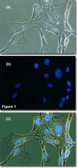
Contrast methods of microscopy including differential interference contrast DIC and phase contrast microscopy can be used in conjunction with WF fluorescence microscopy s. 2D grain structures obtained from such methods can be used to construct 3D RVEs by expanding the structure into the third dimension resulting in columnar formations compare eg. Multiple imaging modes The system offers phase contrast and brightfield label-free imaging fluorescence widefield colorimetric and confocal imaging as well as water immersion imaging as a standard option.
Leica Microsystems exclusively offers Ultra-Contrast 3D illumination. Fluorescence microscopy is a valuable toolbox to study cellular structures and dynamics spanning scales from the single molecule to the live animal. See more details of your sample in less time.
Please read our Terms Conditions and Privacy Policy for information about. Combining WF fluorescence microscopy and contrast techniques can provide valuable information and images of viability and morphology. Phase-contrast imaging is a method of imaging that has a range of different applications.
The spatial resolution that can be achieved with any light-based microscopy is however limited to about 200 nm in the imaging plane and 500 nm along the. Chromatic aberration is compensated to the utmost limit Every single objective is delivered with measured chromatic aberration data and less than 01 µm on-axis chromatic aberration between 405 nm. Using differential-phase-contrast imaging 48 to generate the initial phase leads to an improved initialization 47.
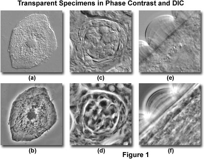
Differential Interference Contrast Comparison Of Phase Contrast And Dic Microscopy Olympus Ls

Differential Interference Contrast Microscopy A Diagram And B Download Scientific Diagram
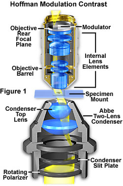
Specialized Microscopy Techniques Hoffman Modulation Contrast Basics Olympus Ls

Phase Contrast Microscopy An Overview Sciencedirect Topics
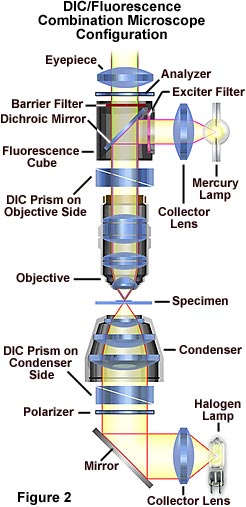
Fluorescence Microscopy Combination Methods With Differential Interference Contrast Dic Olympus Ls

Specialized Microscopy Techniques Contrast In Optical Microscopy Olympus Ls Java Tutorial Paul Dirac Life Science

Phase Contrast Microscopy Combination Methods With Phase Contrast Olympus Ls

Phase Contrast Microscopy By Motic Motic Microscopes

Bursaria Ciliate Protozoan Live Specimen Protozoa Nikon Small World Nikon Small World Specimen Small World
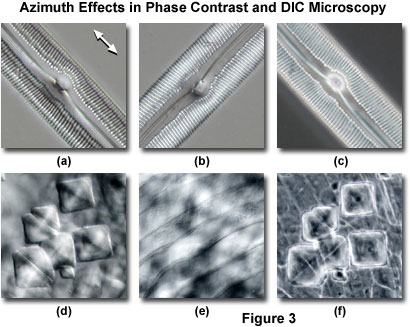
Differential Interference Contrast Comparison Of Phase Contrast And Dic Microscopy Olympus Ls

Phase Contrast Microscopy Combination Methods With Phase Contrast Olympus Ls

Phase Contrast Microscopy An Overview Sciencedirect Topics
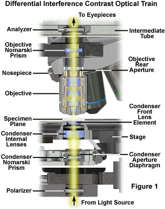
Differential Interference Contrast Dic Microscope Configuration And Alignment Olympus Ls
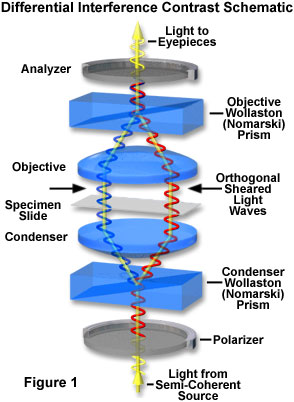
Differential Interference Contrast Introduction Olympus Ls

Differential Interference Contrast Microscopy A Diagram And B Download Scientific Diagram

Keywords Differential Phase Contrast Imaging Keywords Glossary Of Tem Terms Jeol

Phase Contrast Microscopy An Overview Sciencedirect Topics
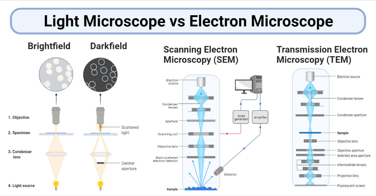
Light Microscope Vs Electron Microscope 36 Major Differences
